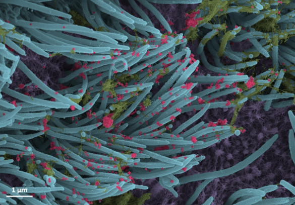
A Close-up of COVID-19 in Lung Cells
Posted on Credit: Ehre Lab, UNC School of Medicine If you or a loved one have come down with SARS-CoV-2, the coronavirus responsible for COVID-19, you know it often takes hold in the respiratory system. This image offers a striking example of exactly what happens to cells in the human airway when this coronavirus infects them. This colorized scanning electron microscope (SEM) image shows SARS-CoV-2-infected human lung cells (purple) covered in hair-like cilia (blue). Those cilia line the inner surface of the airways and help to clear mucus (yellow-green) conta...
Source: nih.gov